|
Cancer - Kidney(구연)
|
(E-081)
|
|
|
종양 윤곽의 불규칙성을 분류하여 국소 신세포암의 종양학적 결과와 예후를 예측할 수 있을까? |
| 부산대학교 의과대학 비뇨기과학 교실 |
| 강병진, 이권경, 박시균, 박지훈, 김경환, 구자윤, 하홍구 |
Introduction
It is known that partial nephrectomy(PN) is standard treatment for localized renal cell carcinoma(RCC). PN is preferred for small renal masses(T1a stage) whenever practicable. Due to the development of surgical techniques, It has recently implemented in T1b-2 renal masses, selectively. This research examines the availability of tumor contour irregularity (TCI) to predict oncologic outcome of clinically localized renal cell carcinoma (RCC).
Materials & methods
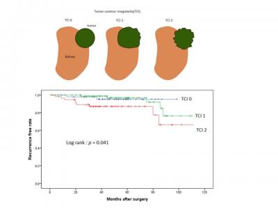
Patients who underwent PN for clinically localized RCC(T1 stage, ≤ 7 cm) with no evident venous or perirenal/sinus fat invasion on imaging were examined. TCI was confirmed by CT/MRI reviewing before surgery, and it was classified into three groups according to tumor shape and contour : TCI 0 (complitely elliptical shape and regular contour), 1 (focally [〈50%] irregular contour), 2 (extensively [≥50%] irregular contour). We conducted chart review and confirmed that there were differences in pathologic outcome and recurrence rate for each TCI groups.
Results
The total number of patients included in this study is 192. Median tumor size was 2.55cm. Male to female ratio was 2.25(n=133) to 1(n=59). TCI was reported as 0/1/2 in 48(25%)/86(44.8%)/58(30.2%) patients, respectively. Upstaging to pathological T3a(n=4) occurred in the TCI 2 with significantly higher probability than in the other groups.(0%/0%/6.9% for TCI 0/1/2, p〈0.05 ). 17 patients (8.9% of total) reported recurrence of cancer during median follow-up of 57 months. There was significantly higher recurrence rate from higher TCI(4.2%/5.8%/17.2%, p〈0.05).
Conclusions
By classification according to TCI, pathologic upstaging and risk of recurrence can be predicted in clinically localized RCC. And TCI could be used as a element of the nephrometry score.
|
 |
|
keywords : renal cell carcinoma, partial nephrectomy, nephrometry |
|